|
 |
 |
 |
 |
|
|
|


Introduction

Absorption is the process whereby toxicants gain entrance into the body. Ingested and inhaled materials are still considered outside the body until they cross the cellular barriers of the gastrointestinal tract or respiratory system. To exert an effect on internal organs it must be absorbed, although local toxicity, such as irritation, may occur.
Absorption varies greatly with specific chemicals and the route of exposure. For skin, oral or respiratory exposure, the exposure dose (outside dose) is only a fraction of the absorbed dose (internal dose). For substances injected or implanted directly into the body, exposure dose is the same as the absorbed or internal dose.
Several factors affect the likelihood that a xenobiotic will be absorbed. The most important are:
 |
 |
 |
|  | route of exposure
|
 |
|  | concentration of the substance at the site of contact
|
 |
|  | chemical and physical properties of the substance
|
 |
The relative roles of concentration and properties of the substance vary with the route of exposure. In some cases, a high percentage of a substance may not be absorbed from one route whereas a low amount may be absorbed via another route. For example, very little DDT powder will penetrate the skin whereas a high percentage will be absorbed when it is swallowed. Due to such route-specific differences in absorption, xenobiotics are often ranked for hazard in accordance with the route of exposure. A substance may be categorized as relatively non-toxic by one route and highly toxic via another route.
The primary routes of exposure by which xenobiotics can gain entry into the body are:
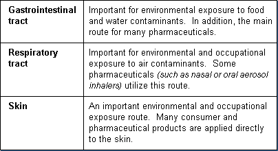
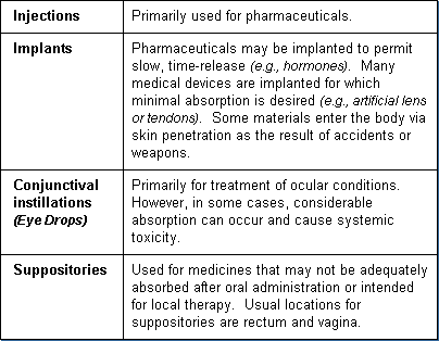
Other routes of exposure - used primarily for specific medical purposes:
For a xenobiotic to enter the body (as well as move within, and leave the body) it must pass across cell membranes (cell walls). Cell membranes are formidable barriers and a major body defense that prevents foreign invaders or substances from gaining entry into body tissues. Normally, cells in solid tissues (such as skin or mucous membranes of the lung or intestine) are so tightly compacted that substances can not pass between them. This requires that the xenobiotic have the ability to penetrate cell membranes. It must cross several membranes in order to go from one area of the body to another.
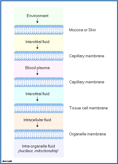
In essence, for a substance to move through one cell requires that it first move across the cell membrane into the cell, pass across the cell, and then cross the cell membrane again in order to leave the cell. This is true whether the cells are in the skin, the lining of a blood vessel, or an internal organ (e.g., liver). In many cases, in order for a substance to reach the site of toxic action, it must pass through several membrane barriers.
As illustrated in the diagram below, a foreign chemical will pass through several membranes before it comes into contact with and can damage the nucleus of a liver cell.
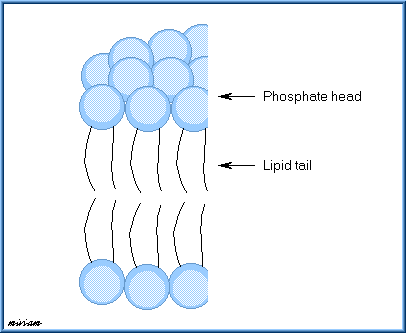
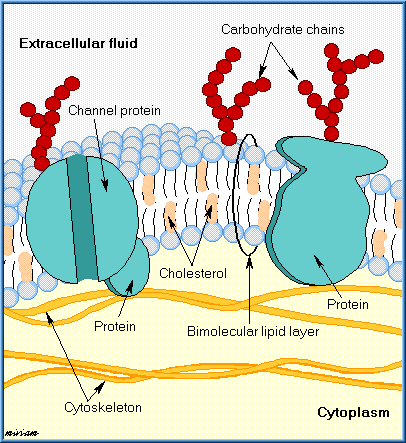
Cell membranes (often referred to as plasma membranes) surround all body cells and are basically similar in structure. They consist of two layers of phospholipid molecules arranged like a sandwich, referred to as a "phospholipid bilayer". Each phospholipid molecule consists of a phosphate head and a lipid tail. The phosphate head is polar, that is it is hydrophilic (attracted to water). In contrast, the lipid tail is lipophilic (attracted to lipid-soluble substances).
The two phospholipid layers are oriented on opposing sides of the membrane so that they are approximate mirror images of each other. The polar heads face outward and the lipid tails inward in the membrane sandwich.
The cell membrane is tightly packed with these phospholipid molecules interspersed with various proteins and cholesterol molecules. Some proteins span across the entire membrane providing for the formation of aqueous channels or pores.
A typical cell membrane structure is illustrated below.
Some toxicants move across a membrane barrier with relative ease while others find it difficult or impossible. Those that can cross the membrane, do so by one of two general methods, passive transfer or facilitated transport.
Passive transfer consists of simple diffusion (or osmotic filtration) and is "passive" in that there is no cellular energy or assistance required.
Some toxicants can not simply diffuse across the membrane but require assistance or facilitated by specialized transport mechanisms. The primary types of specialized transport mechanisms are:
 |
 |
 |
|  | facilitated diffusion
|
 |
|  | active transport
|
 |
|  | endocytosis (phagocytosis and pinocytosis)
|
 |
Passive transfer is the most common way that xenobiotics cross cell membranes. Two factors determine the rate of passive transfer:
 |
 |
 |
|  | difference in concentrations of the substance on opposite sides of the membrane (substance moves from a region of high concentration to one having a lower concentration. Diffusion will continue until the concentration is equal on both sides of the membrane)
|
 |
|  | ability of the substance to move either through the small pores in the membrane or the lipophilic interior of the membrane
|
 |
Properties of the chemical substance that affect its' ability for passive transfer are:
 |
 |
 |
|  | lipid solubility
|
 |
|  | molecular size
|
 |
|  | degree of ionization
|
 |
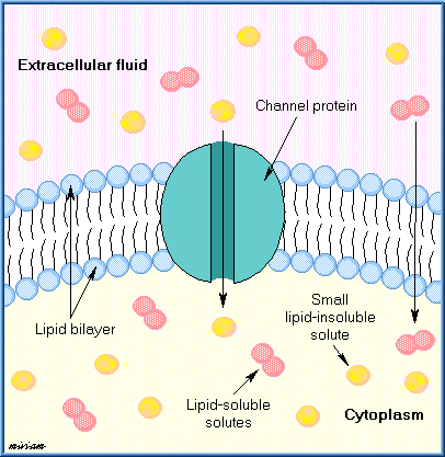
Substances with high lipid solubility readily diffuse through the phospholipid membrane. Small water-soluble molecules can pass across a membrane through the aqueous pores, along with normal intracellular water flow.
Large water-soluble molecules usually can not make it through the small pores, although some may diffuse through the lipid portion of the membrane, but at a slow rate. In general, highly ionized chemicals have low lipid solubility and pass with difficulty through the lipid membrane.
Most aqueous pores are about 4Å in size and allow chemicals of molecular weight 100-200 to pass through. Exceptions are membranes of capillaries and kidney glomeruli which have relatively large pores (about 40Å) that allow molecules up to a molecular weight of about 50,000 (molecules slightly smaller than albumen which has a molecular weight of 60,000) to pass through.
The illustration below demonstrates the passive diffusion and filtration of xenobiotics through a typical cell membrane.
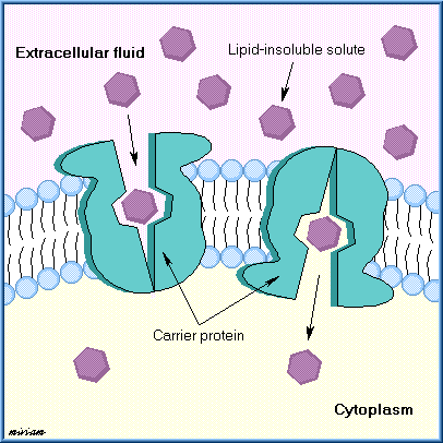
Facilitated diffusion is similar to simple diffusion in that it does not require energy and follows a concentration gradient. The difference is that it is a carrier-mediated transport mechanism. The results are similar to passive transport but faster and capable of moving larger molecules that have difficulty diffusing through the membrane without a carrier. Examples are the transport of sugar and amino acids into RBCs and the CNS.
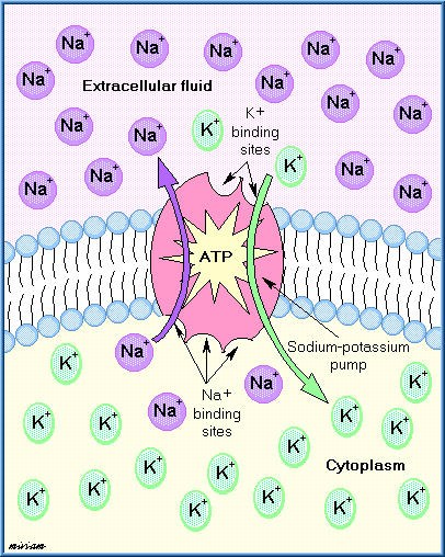
Some substances are unable to move with diffusion, unable to dissolve in the lipid layer, and are too large to pass through the aqueous channels. For some of these substances, active transport processes exist in which movement through the membrane may be against the concentration gradient, that is, from low to higher concentrations. Cellular energy from adenosine triphosphate (ADP) is required in order to accomplish this. The transported substance can move from one side of the membrane to the other side by this energy process. Active transport is important in the transport of xenobiotics into the liver, kidney, and central nervous system and for maintenance of electrolyte and nutrient balance.
In the following figure, the sodium and potassium ions are moving against concentration gradient with the help of the ADP sodium-potassium pump.
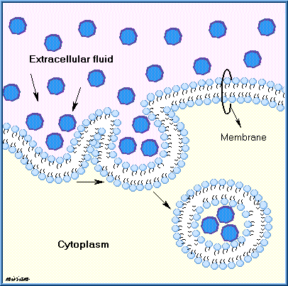
Many large molecules and particles can not enter cells via passive or active mechanisms. However, some may still enter by a process known as endocytosis.
In endocytosis, the cell surrounds the substance with a section of its cell wall. This engulfed substance and section of membrane then separates from the membrane and moves into the interior of the cell. The two main forms of endocytosis are phagocytosis and pinocytosis.
In phagocytosis (cell eating), large particles suspended in the extracellular fluid are engulfed and either transported into cells or are destroyed within the cell. This is a very important process for lung phagocytes and certain liver and spleen cells. Pinocytosis (cell drinking) is a similar process but involves the engulfing of liquids or very small particles that are in suspension within the extracellular fluid.
The illustration below demonstrates endocytosis membrane transport.

  
|
|
|
|


